Imaging of the Charcot foot everything you need to know you will find in this open access review article by A. Rosskopf:
-
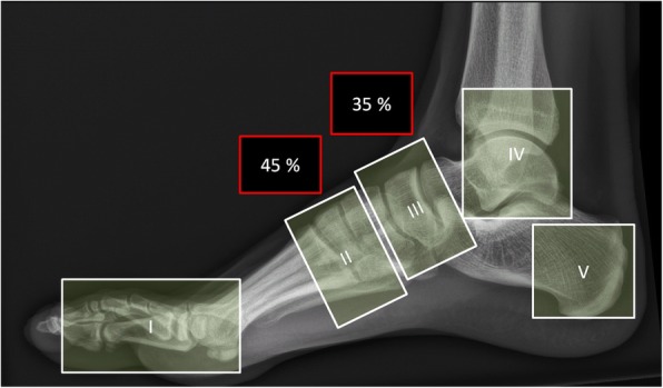
X-rays may be normal during early stage
-
MRI should cover the entire foot
-
MRI can be used for early diagnosis, monitoring of disease activity and complications
-
Acute MRI findings include bone marrow edema, soft tissue edema, and subchondral fractures
-
Chronic MRI findings include subchondral cysts, joint destructions, joint effusion, and bony proliferations