3D-printed anatomic models of the knee for evaluation of patellofemoral dysplasia in comparison to standard radiographs and computed tomography
Benjamin Fritz, Sandro F. Fucentese, Stefan M. Zimmermann, Philippe M. Tscholl, Reto Sutter, Christian W.A. Pfirrmann
European Journal of Radiology Published:April 17, 2020
- 3D models can increase accuracy for detection of patellofemoral dysplasia.
- For Dejour classification, 3D models and radiographs/CT show similar performances.
- Inter-reader reliabilities are similar of 3D model and radiograph/CT assessments.
Purpose
To evaluate 3D-printed anatomic models of the distal femur and patella for diagnosis and classification of patellofemoral dysplasia in comparison to conventional radiographs (CR) and CT.
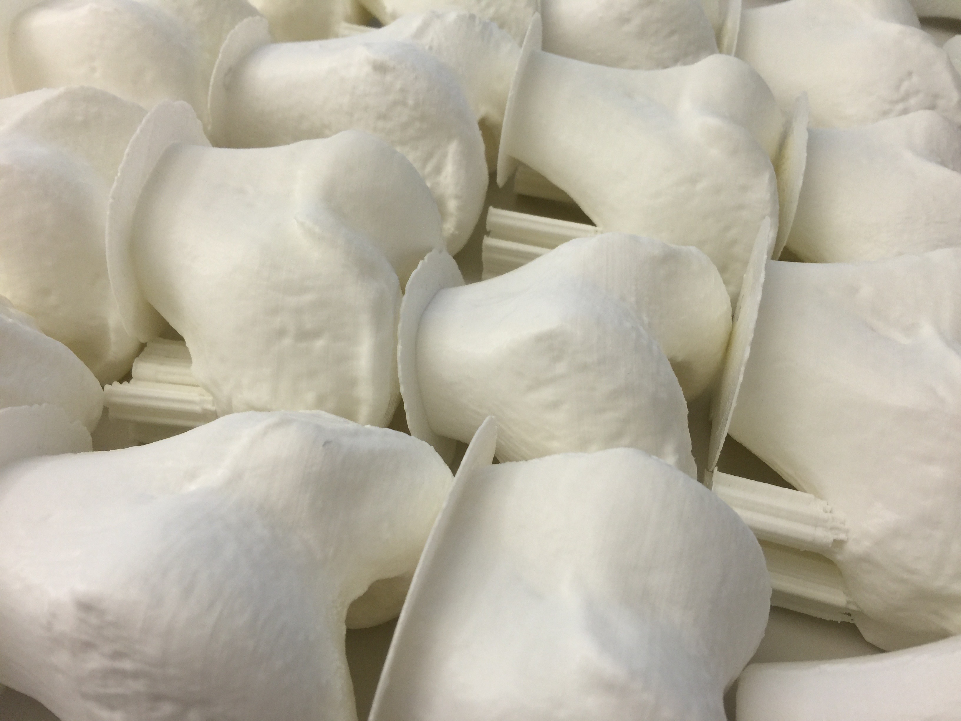
Method: Following local ethics committee approval, CT-datasets of 50 patients were segmented and 3D-anatomic models of the distal femur and patella were printed. An expert panel reviewed CR, CT, 3D-models and patient history and classified the femoral trochleas into normal or Dejour type A–D and the patellas into Wiberg type A–C, which served as the standard of reference. The same classifications were performed by two readers independently, first based on 3D-models and after 3 weeks based on CR and CT. Descriptive statistics, ROC-analysis and inter-reader reliability were performed.
Results: Trochlear dysplasia was present in 28/50 patients. Evaluations of 3D-models vs. CR/CT for trochlear dysplasia showed a sensitivity/specificity of 89.3 %/100 % vs. 96.4 %/68.2 % for reader 1 and 96.4 %/100 % vs. 96.4 %/90.9 % for reader 2, and an area under the curve of 0.946 vs. 0.823 for reader 1 (p = 0.029) and 0.982 vs. 0.937 for reader 2 (p = 0.147). Evaluations of 3D-models vs. CR/CT for the Dejour classification showed a sensitivity/specificity of 32.1 %/100 % vs. 57.1 %/68.2 % for reader 1 and 46.4 %/100 % vs. 50 %/90.9 % for reader 2 without significant differences. No significant differences existed for Wiberg-classification (50–66 % exact matches) or inter-reader reliabilities between 3D-models and CR/CT for all assessments (Kappa 0.428–0.92).
Conclusion: In comparison to radiographs and CT, 3D-models achieve similar diagnostic accuracy for detection of patellofemoral dysplasia and have the potential to improve diagnosis for less experienced physicians.